The critical period for the brain to learn to make sense of visual input has been debated by neuroscientists for decades, with the consensus resting on the ages of 6 and 7. The study was published in PNAS.
Professor Pawan Sinha of MIT has recently shown that there is more nuance to the picture than that. He has found that older children can learn visual tasks like face recognition, object separation, and motion perception in a number of studies of Indian children who had surgery to remove congenital cataracts after the age of 7.
The researchers have found in a new study that these patients’ brains undergo anatomical changes after they regain sight. Researchers also noted improvements in patients’ eyesight, which they attribute in part to these alterations in the white matter of the brain.
The results provide more evidence that the window of brain plasticity is much wider than previously believed, at least for some visual tasks.
The goal of the current study was to see if the researchers could find any anatomical changes in the brain that might explain the observed behavioral changes in treated children. Participants’ ages ranged from 7 to 17, and they were scanned multiple times after having congenital cataracts surgically removed.
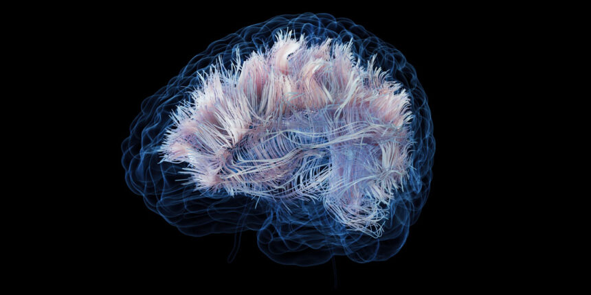
The team used diffusion tensor imaging, a subset of MRI used specifically for studying brain anatomy changes. White matter (groups of nerve fibers that connect different parts of the brain) organization changes can be seen with this imaging technique.
Mean diffusivity, a measure of the ease with which water molecules can move, and fractional anisotropy, a measure of the relative difficulty of water moving in different directions, are the two measurements produced by diffusion tensor imaging, which follows the motion of hydrogen nuclei in water molecules.
An increase in fractional anisotropy indicates that water molecules are more constrained due to the orientation of nerve fibers in the white matter, according to researchers.


