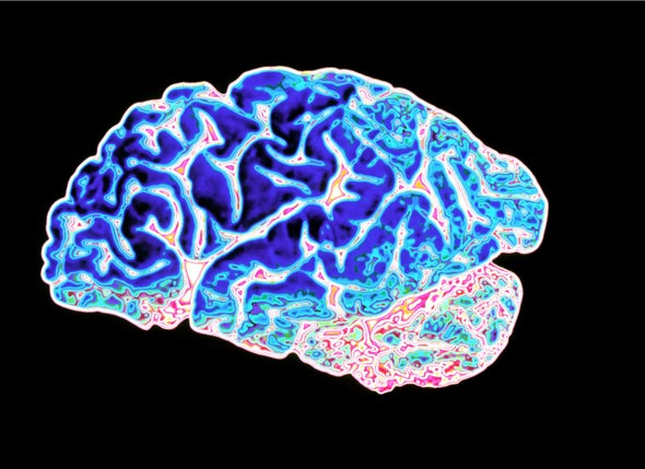
With the use of tau-positron emission tomography scans of more than 1,600 participants, a research group at Lund University established four distinct subtypes of Alzheimer’s disease, based on tau pathology.
Released in Nature Medicine, a peer-reviewed journal, the study aimed at highlighting how the tau protein spreads in the brain in distinct patterns, often producing different symptoms.
According to the study, four varients were established.
In the first variant, the tau protein spreads primarily within the temporal lobe, impacting memory, while variant two spreads in the rest of the cerebral cortex. Heightened difficulties with executive function are evident in this regard.
The third variant involves the accumulation of tau in the visual cortex, in which the visuospatial processing of sensory impressions in the brain is impacted. As part of variant four, tau spreads in the left hemisphere of the brain.
From the findings: “The subtypes presented with distinct demographic and cognitive profiles and differing longitudinal outcomes. Additionally, network diffusion models implied that pathology originates and spreads through distinct corticolimbic networks in the different subtypes.”
“Together, our results suggest that variation in tau pathology is common and systematic, perhaps warranting a re-examination of the notion of ‘typical AD’ and a revisiting of tau pathological staging.”


