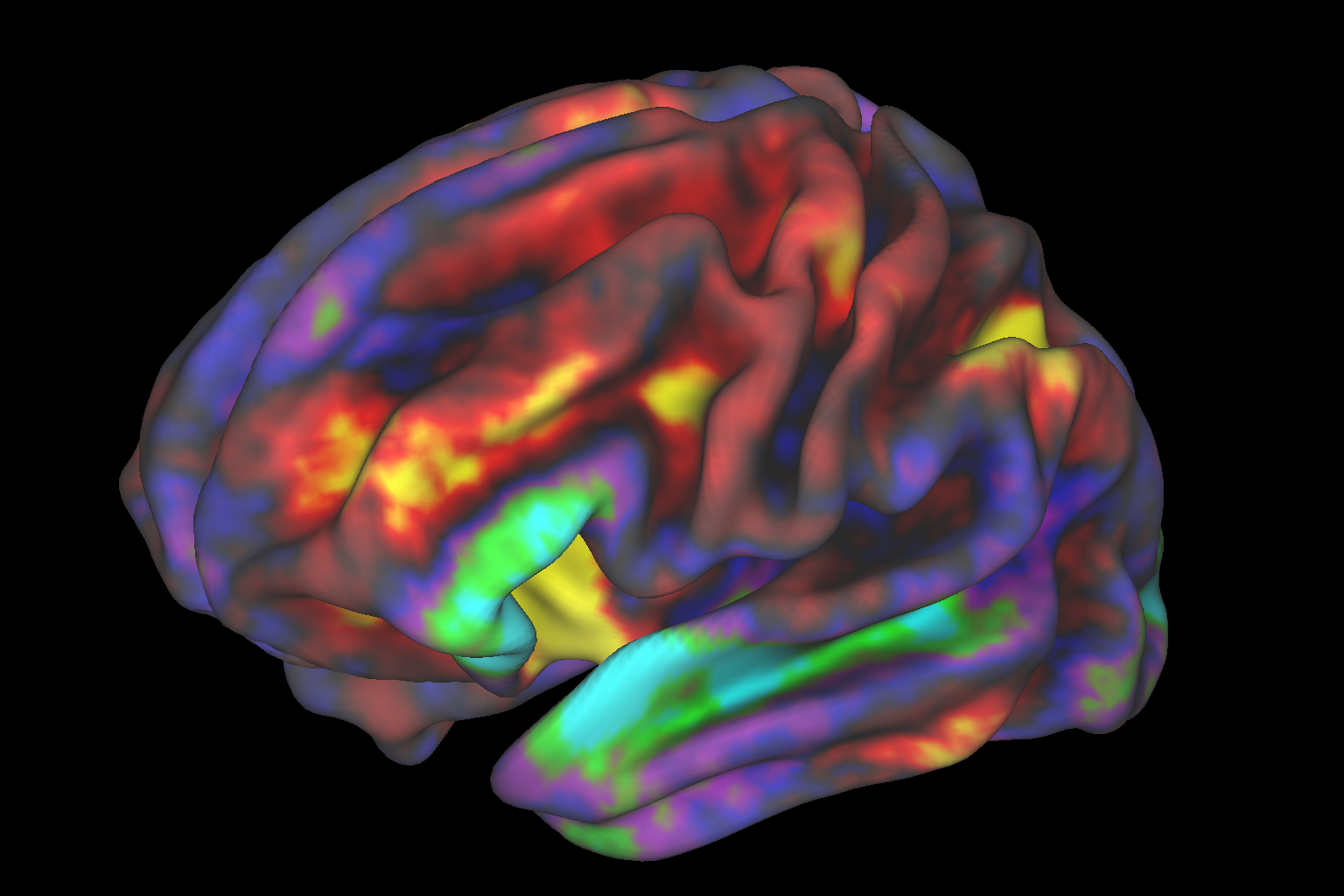
A team of researchers at Yale University were able to reduce, for the first time, the severity of symptoms associated with Tourette syndrome (TS) by using real-time brain imaging. The findings appeared in Biological Psychiatry.
“It is a non-invasive, neuroscience-based intervention for training human brain function towards healthier patterns,” said Michelle Hampson, senior author of the study.
The team examined 21 adolescents, aged 11 to 19, with particular features like repetitive movements, or tics, utilizing the Yale Global Tic Severity Scale, as part of a double-blind, randomized, crossover study. The participants were instructed to initiate activity in the supplementary motor area of the brain, then monitor the function and activity in real-time using brain imaging. The supplementary motor area is thought to play a major role in the behavioral exhibitions of Tourette syndrome.
Researchers trained the participants with real-time functional magnetic resonance imaging neurofeedback (rt-fMRI-NF) which allowed for the monitoring of function and activity in the brain. The technique has previously been utilized for other psychiatric conditions, like depression, but never for this neurodevelopmental disorder.
The findings showed significant improvements in tics and repetitive movements after neurofeedback compared to their counterpart. “This first randomized controlled trial of rt-fMRI neurofeedback in adolescents with TS suggests that this neurofeedback intervention may be helpful for improving tic symptoms,” researchers say.
“This is the first study of its kind showing that rt-fMRI-NF has potential as a treatment for Tourette Syndrome.”


